News
Search All News
Top Stories
-
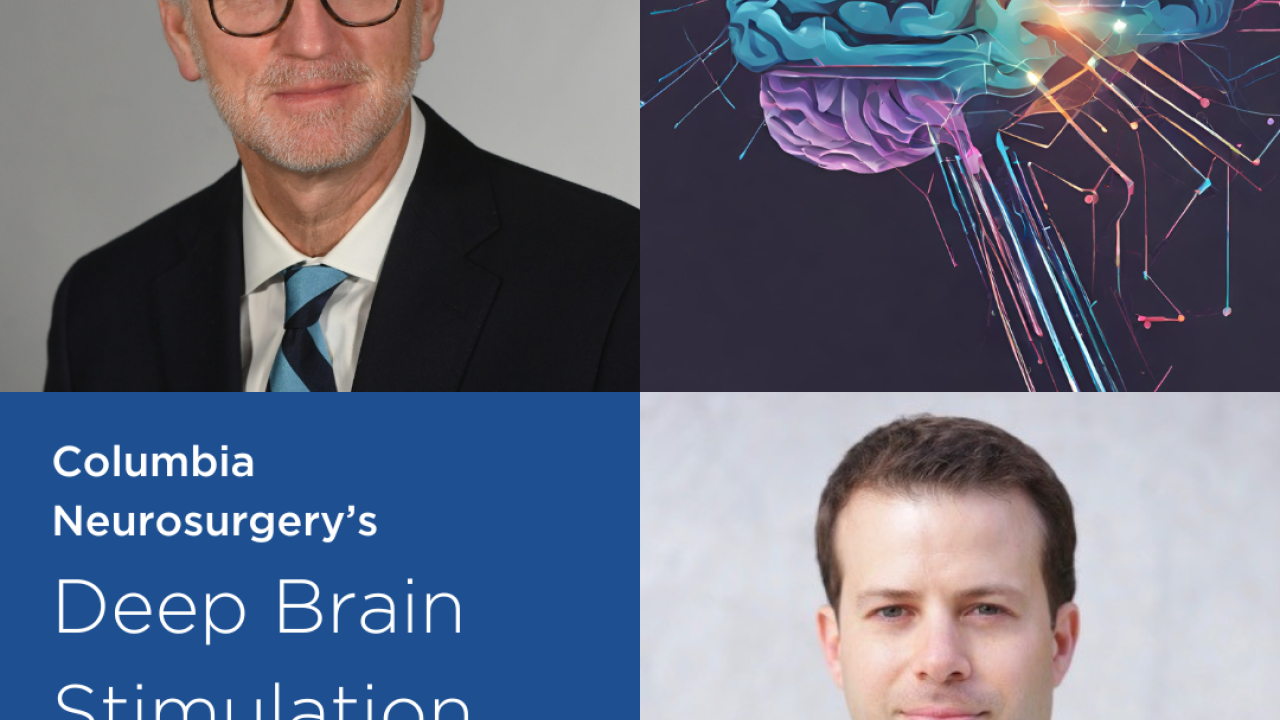
Columbia Neurosurgery is now one of the few places in the world where patients can benefit from Asleep DBS, meaning patients are sedated throughout the procedure.
-
Columbia Neurosurgery shares Dr. Karin Marie Muraszko received the VP&S Alumni Association’s Virginia Kneeland Frantz ’22 Award for Distinguished Women in Medicine.
-
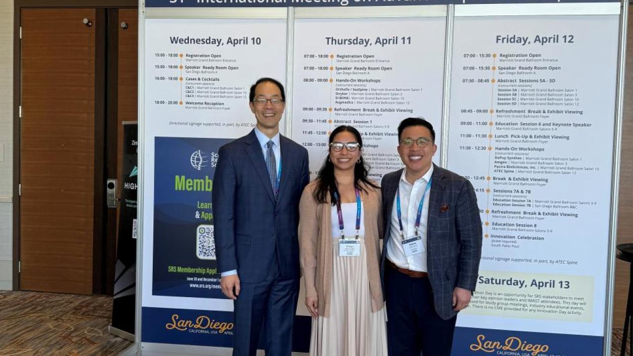
Dr. Dean Chou, Dr. Andrew Chan and Spine Clinical Research Manager Vivian Le traveled to San Diego for the 31st International Meeting on Advanced Spine Techniques (IMAST).
-
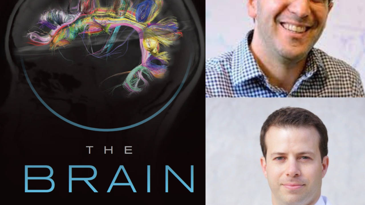
NIH BRAIN Initiative awarded Josh Jacobs and Brett Youngerman, MD, a new award for $3.1million over 3 years with potential to expand to over $5 million over 5 years.