Neurosurgery and Radiology: The Role of the Neuroradiologist
Spotlight on Skull Base: Head and Neck
The role of the neuroradiologist is critical for neurosurgical patient care. Columbia Neurosurgery works with some of the most experienced and specialized in the field to provide a comprehensive and interdisciplinary approach to care. A neuroradiologist obtains further training in treating the spine, head, neck and the central and peripheral nervous systems. The Neurological Institute of New York here at CUIMC was the first hospital in the western hemisphere dedicated to neurosurgery and radiology. Through the last 100 years, the team has evolved into a highly specialized model with key radiology team members serving each division.
Specifically, for skull base cases involving the head and neck members of the Radiology faculty, Dr. Akinrinola Famuyide, Dr. Pamela Nguyen, and Dr. William Gomes closely partner with Neurosurgery's Dr. Raymond Sekula and Dr. Brett Youngerman.
Says Dr. Famuyide, "Working at Columbia, I'm struck by the number of patient care conferences we have that bring a multidisciplinary team together. You end up with a team of multiple doctors and care providers discussing your care and producing the best treatment plan.
Dr. Nguyen remarked, "The scope of pathology here at Columbia requires a well-coordinated multidisciplinary approach, and we are very lucky to have such amazing colleagues in each department."
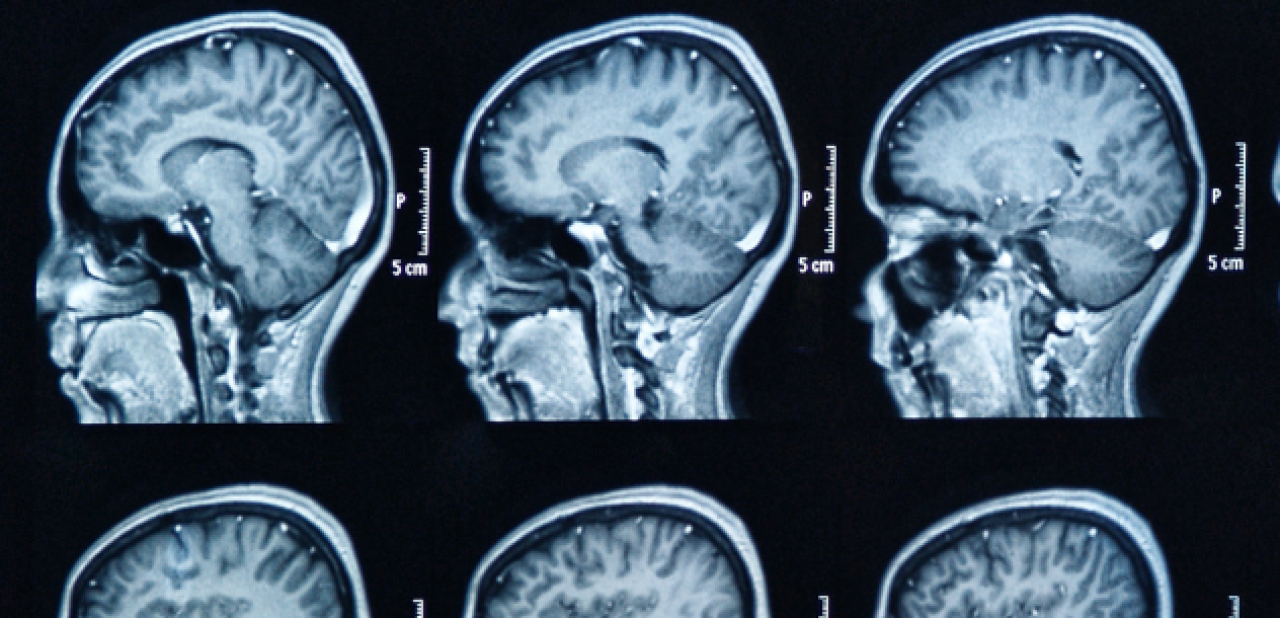
Says Dr. Raymond Sekula, "When working with our neuroradiologists, I am energized by the fact that we have developed the highest resolution MRI and other protocols available in the United States, which has dramatically transformed our ability to care for patients with hard to reach skull base tumors, including meningiomas, acoustic neuromas, and other disorders as well as cranial neuralgias, such as trigeminal neuralgia, hemifacial spasm, and glossopharyngeal neuralgia. With patients traveling each week from across the U.S. for our services, we provide top-of-class neurosurgical care."
Concerning functional brain mapping, Dr. Brett Youngerman shared, "We are deploying the latest advanced imaging techniques for targeting difficult-to-see structures in the brain and avoiding function critical sites. Advanced structural imaging techniques and diffusion tensor imaging, also known as tractography, allow us to target millimeter-sized structures deep in the brain that were previously not visible but are essential for outcomes of neuromodulation therapy for drug-resistant epilepsy and Parkinson's disease. Advanced imaging also allows us to identify the onset of seizures in drug-resistant epilepsy. Other functional imaging techniques allow us to protect critical functions like speech, motor, and vision."
Most recently, in Radiology and AI, Columbia Medical Center announced its new Center for Innovation in Imaging Biomarkers and Integrated Diagnostics (CIMBID), dedicated to developing and integrating quantitative imaging and non-imaging biomarkers for disease prediction, particularly in cancer. Despina Kontos, PhD, a computer scientist with expertise in artificial intelligence, machine learning, and big data analytics for multi-modality imaging data, leads the center.

